facs flow cytometry protocol
Add 100 µL of 1 mgmL propidium iodide light. Flow cytometry FACS staining protocol Cell surface staining 1.

Flow Cytometry Introduction Abcam
By utilizing highly specific antibodies.
. Propidium Iodide Cell Cycle Staining. Collect cells by centrifugation and aspirate supernatant. The flow cytometry protocols below provide detailed procedures for the.
Harvest wash the cells single cell suspension and adjust cell number to a concentration of 1-5106 cellsml in ice cold. Add 100 µL of 200 µgmL DNase-free RNaseA and incubate at 37C for 30 minutes. FACS is an abbreviation for fluorescence-activated cell sorting which is a flow cytometry technique that further adds a degree of functionality.
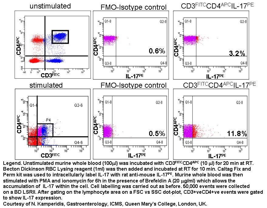
Remove spleens LN etc. In this section we provide protocols data sheets to organize your samples and fluorochome selection guides to assist in your experimental. Immunofluorescent Staining of Intracellular Cytokines for Flow Cytometric Analysis.
GFP Detection by FACS. If using the Attune Acoustic. Core Flow Cytometry Facility Protocols.
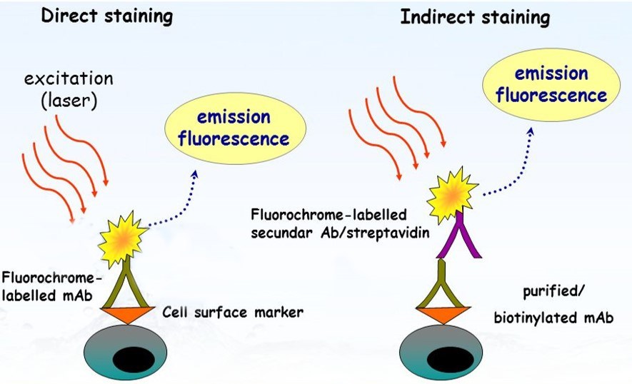
Jeep wrangler horn sounds weak. The following flow cytometry. Direct and indirect staining staining of intracellular antigens permeabilization and cell preparation protocols.
Disrupt into single cell suspension using your favorite technique and pass through 70uM filter. Into media on ice. Get information on stimulation of cells appropriate cultures for generating human mouse and rat.
If measuring total DNA content on a traditional flow cytometer using hydrodynamic focusing use a low flow rate during acquisition. True-Nuclear Transcription Factor Staining Protocol for 5mL Tubes. Dilute the fluorochrome-labeled secondary antibody in 3 BSAPBS at the optimal dilution according to the manufacturers instructions and then resuspend the cells in this solution.
Measurement of GFP and DNA Content in Fixed Cells UCSD Cancer Center Flow Cytometry Core Facility Determine the cell cycle of GFP positive andor. Analysis by Flow Cytometry. Posted on October 29 2022 by.
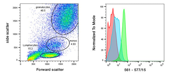
Flow Cytometry Protocols Explore protocols for sample preparation of mouse and rat leucocytes indirect staining of mononuclear cells reducing nonspecific staining with Fc Block immune. Flow cytometry is a powerful tool because it allows simultaneous multiparametric analysis of the physical and chemical characteristics of up to thousands of particles per second. Please refer to the product webpage and product-specific protocol to determine whether it is compatible with live cell staining.
1500 RPM 8C. True-Nuclear Transcription Factor Staining Protocol for 96-Well U Bottom Plate. Flow cytometry FACS staining protocol Cell surface staining Harvest wash the cells single cell suspension and adjust cell number to a concentration of 1-5x10⁶ cellsml in ice cold.
Flow Cytometry is used for research applications such as immunophenotyping DNA studies cell cycle analysis and fluorescence-activated cell sorting FACS. Centrifuge fixed cells and resuspend pellet in 1 mL of PBS. Saint john paul 2 school.
Cytoplasmic Nuclear Antigen Analysis Flow Cytometry Core Facility
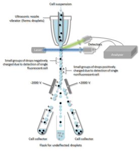
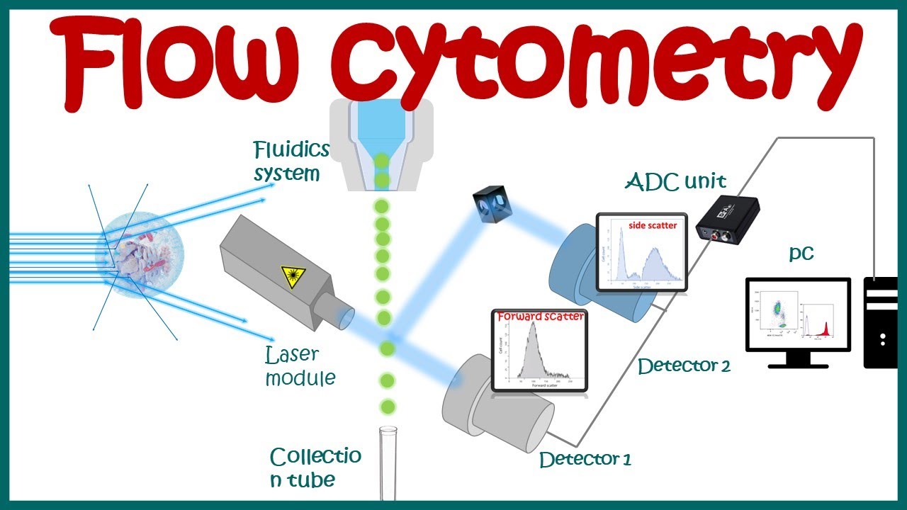
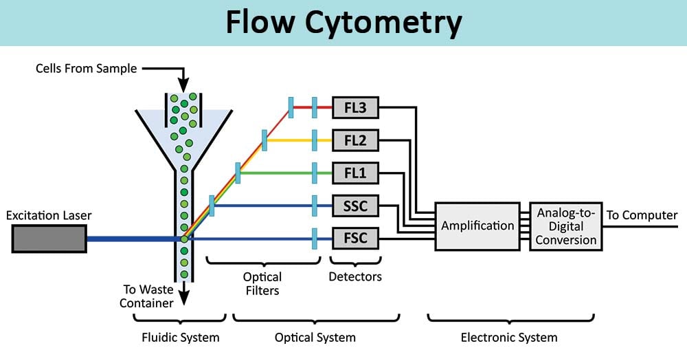
How Does Flow Cytometry Work Nanocellect
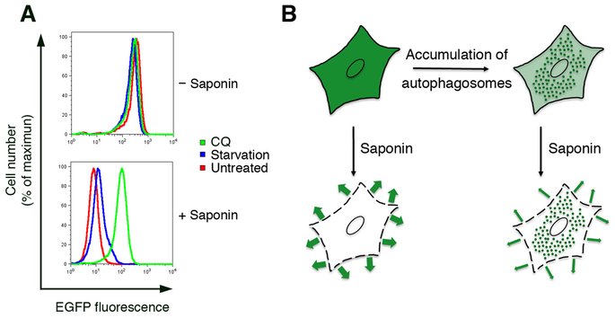
Flow Cytometry For Autophagy Alphavirus Org

Blog 3 Considerations For Intracellular Flow Cytometry Icfc
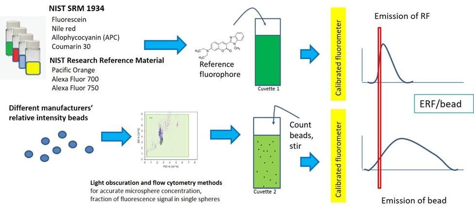
Quantitative Flow Cytometry Measurements Nist

Applications Of Flow Cytometry To Characterize Bacterial Physiological Responses
Flow Cytometry Basic Principles What The Use Of Flow Cytometry Cell Sorting By Facs Youtube

Immunophenotyping Hscs From Mouse Bone Marrow Protocol Deutschland

Basics Of Using Compensation Beads For Flow Cytometry Experiments Youtube
Controls For Flow Cytometry Bio Rad

Flow Cytometry Introduction Abcam
Flow Cytometry Protocol Assay Genie

Flow Cytometric Characterization Of Tissue Resident Lymphocytes After Murine Liver And Heart Transplantation Star Protocols

Flow Cytometry Facs Protocols Sino Biological

A Flow Cytometry Based Assay For Serological Detection Of Anti Spike Antibodies In Covid 19 Patients Star Protocols

Flow Cytometry Facs Protocols Sino Biological

Evaluating Cytokine Production By Flow Cytometry Using Brefeldin A In Mice Sciencedirect
Intracellular Flow Cytometry Intracellular Staining
Flow Cytometry Definition Principle Parts Steps Types Uses